Dental CBCT Scan
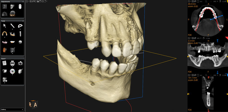
Dental Cone Beam CT. Dental cone beam computed tomography (CT) is a special type of x-ray equipment used when regular dental or facial x-rays are not sufficient. Your dentist may use this technology to produce three dimensional (3-D) images of your teeth, soft tissues, nerve pathways and bone in a single scan.
Available from £140
What is a Dental CBCT Scan?
A Dental Cone Beam CT scan offers a highly detailed view of your oral structures, allowing your dentist to examine areas that cannot be seen clearly with standard x-rays. The 3D imaging provides precise information about your teeth, jawbone, and surrounding tissues, helping to plan treatments such as dental implants, extractions, orthodontics, and root canal therapy with greater accuracy and confidence.
This advanced technology uses a focused cone-shaped beam to capture comprehensive images in a single scan, significantly reducing the need for multiple x-rays. It ensures minimal radiation exposure while delivering exceptional diagnostic detail, enabling your dentist to assess your oral health and structure with precision and care.

Frequently Asked Questions
Dental cone beam computed tomography (CT) is a special type of x-ray machine used in situations where regular dental or facial x-rays are not sufficient. It is not used routinely because the radiation exposure from this scanner is significantly more than regular dental x-rays. This type of CT scanner uses a special type of technology to generate three dimensional (3-D) images of dental structures, soft tissues, nerve paths and bone in the craniofacial region in a single scan. Images obtained with cone beam CT allow for more precise treatment planning.
Cone beam CT is not the same as conventional CT. However, dental cone beam CT can be used to produce images that are similar to those produced by conventional CT imaging.
With cone beam CT, an x-ray beam in the shape of a cone is moved around the patient to produce a large number of images, also called views. CT scans and cone beam CT both produce high-quality images.
Dental cone beam CT was developed as a means of producing similar types of images but with a much smaller and less expensive machine that could be placed in an outpatient office.
Cone beam CT provides detailed images of the bone and is performed to evaluate diseases of the jaw, dentition, bony structures of the face, nasal cavity and sinuses. It does not provide the full diagnostic information available with conventional CT, particularly in evaluation of soft tissue structures such as muscles, lymph nodes, glands and nerves. However, cone beam CT has the advantage of lower radiation exposure compared to conventional CT.
Dental cone beam CT is commonly used for treatment planning of orthodontic issues. It is also useful for more complex cases that involve:
- Surgical planning for impacted teeth.
- Diagnosing temporomandibular joint disorder (TMJ).
- Accurate placement of dental implants.
- Evaluation of the jaw, sinuses, nerve canals and nasal cavity.
- Detecting, measuring and treating jaw tumours.
- Determining bone structure and tooth orientation.
- Locating the origin of pain or pathology.
- Cephalometric analysis.
- Reconstructive surgery.
A cone beam CT examination requires no special preparation.
Prior to the examination, you may be asked to remove anything that may interfere with the imaging, including metal objects, such as jewellery, eyeglasses, hairpins and hearing aids. Although removable dental work may need to be removed, it is advisable to bring these to your examination, as your dentist or oral surgeon may need to examine these as well.
Women should always inform their dentist or oral surgeon if there is any possibility that they are pregnant.
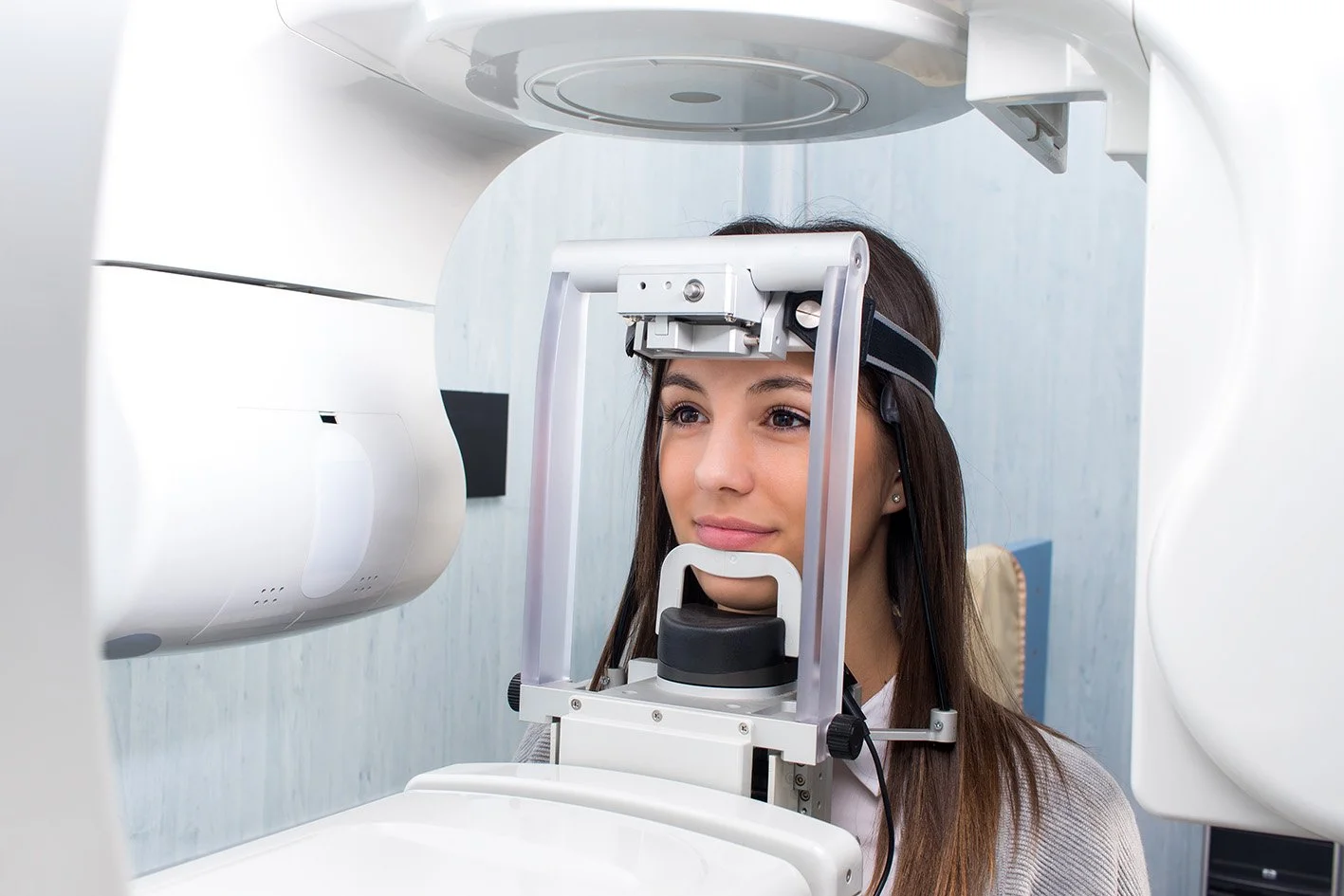
Cone beam CT scanners are typically square-shaped machines designed for patients to remain upright throughout the scan. They feature a rotating C-arm, which holds the x-ray source and detector, and moves around the patient to capture detailed 3D images. Unlike other scanners, these machines do not include a chair or table, so patients must stand for the entire examination.
During a cone beam CT examination, the C-arm or gantry rotates around the head in a complete 360-degree rotation while capturing multiple images from different angles that are reconstructed to create a single 3-D image.
The x-ray source and detector are mounted on opposite sides of the revolving C-arm or gantry and rotate in unison. In a single rotation, the detector can generate anywhere between 150 to 200 high resolution two-dimensional (2-D) images, which are then digitally combined to form a 3-D image that can provide your dentist or oral surgeon with valuable information about your oral and craniofacial health.
During the scan you will be standing holding handles for stability that are either side of the scanner. Your dentist or oral surgeon will position you so that the area of interest is centred in the beam. You will be asked to remain very still while the x-ray source and detector revolve around you for a 360-degree rotation or less. This typically can take between 20 to 40 seconds for a complete volume, also called a full mouth x-ray, in which the entire mouth and dental structures are imaged, and less than 10 seconds for a regional scan that focuses on a specific area of the maxilla or mandible.
You will not experience any pain during a cone beam CT exam, and you will be able to return to your normal activities once the exam is complete.
The images will be reviewed by your dentist, oral surgeon, or radiologist. If you are a patient at our clinic, we will discuss the results with you. If you were referred to us, please note that we do not provide a clinical opinion. You will receive a CD copy of the scan to take to your referring dentist, who can then review the results with you.
Benefits
- The focused x-ray beam reduces scatter radiation, resulting in better image quality.
- A single scan produces a wide variety of views and angles that can be manipulated to provide a more complete evaluation.
- Cone beam CT scans provide more information that conventional dental x-ray, allowing for more precise treatment planning.
- CT scanning is painless, non-invasive and accurate.
- A major advantage of CT is its ability to image bone and soft tissue at the same time.
- No radiation remains in a patient’s body after a CT examination.
- X-rays used in CT scans should have no immediate side effects.